Dental Technology
Intraoral Technology
Many patients, especially younger patients, are very familiar with the latest technology and are more comfortable with the high-tech practice. Computers and TV screens are their primary method of information processing. Dr. Caron and Dr. Kenney utilize intraoral camera technology that helps enhance your understanding of your diagnosis. An intraoral camera is a very small camera. In some cases, an intraoral camera is just a few millimeters long. An intraoral camera allows our practice to view clear, precise images of your mouth, teeth, and gums in order for us to accurately make a diagnosis. With clear, defined, enlarged images, you see details that may be missed by standard mirror examinations. This can mean faster diagnosis with less chair-time for you!
Intraoral cameras also enable our practice to save your images in our office computer to provide a permanent record of treatments. These treatments can be printed for you, other specialists, and your lab or insurance companies.
Precision Dentistry
When you seek care at our office, you are assured that Dr. Caron and Dr. Kenney and their staff utilize the latest in technology to enhance the quality and fit for your dental care.
Dentistry is micro-surgery. Using a similar microscope that an ophthalmologist uses enables us to create dental restorations with incredibility precise fit and finish. You just can't fulfill that level of care with the naked eye.
In addition to allowing precise, close-up work, the microscope directs a beam of light directly on the teeth, minimizing glare to you.
For the most precise aspects of restorative procedures, we use electric handpieces. This results in extraordinarily precise interfaces between your tooth and your new restoration (crown, veneer, or filling). This will bring more comfort to you as well. With a more precise tool, there is less vibration and less noise!
Digital Imaging
Dr. Caron and Dr. Kenney choose carefully which and when radiographs are taken. There are many guidelines that we follow. Radiographs allow us to see everything we cannot see with our own eyes. Radiographs enable us to detect cavities in between your teeth, determine bone level, and health of bone. We can also examine the roots and nerves of teeth, diagnose lesions, such as cysts or tumors, as well as assess damage when trauma occurs. Dental radiographs are invaluable aids in diagnosing, treating, and maintaining dental health. Exposure time for dental radiographs is extremely minimal.
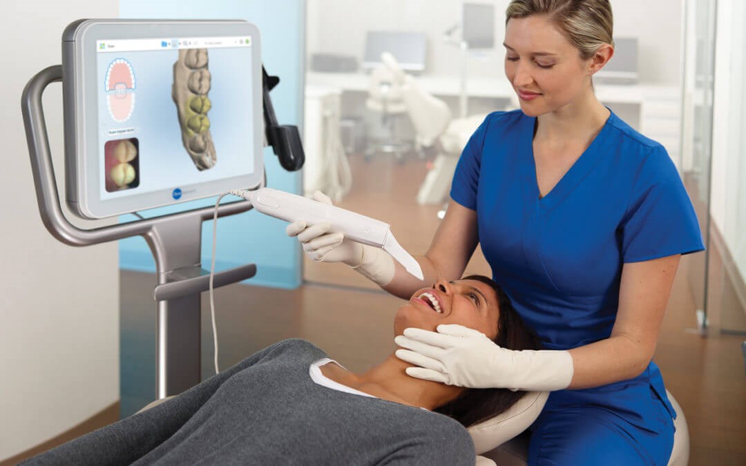
Doctors Caron and Kenney utilize digital imaging technologies within the office, including the iTero intraoral scanner. With digital imaging, exposure time is about 90 percent less when compared to traditional radiographs. Digital imaging can also help us retrieve valuable diagnostic information. We may be able to see cavities better. The advantages of digital imaging enable us to not only store patient images but also enables us to quickly transfer them to appropriate specialists or insurance companies.
Digital X-rays
Digital x-rays offer more precision since we view the image on a computer monitor instead of holding up a 35mm film up to the light. Digital x-rays result in one-sixth of the radiation exposure to you.
STA SingleTooth Anesthesia System
The STA SingleTooth Anesthesia System is a computer-assisted system for local anesthesia. The instrument carefully guides our doctors as they perform injections. Patients benefit from our technology with less discomfort and will not have collateral numbness. Drs. Caron, Kenney, and Russell are delighted to offer the STA system to our dental clients.
Sprint Ray Pro 3D Printer
Tupelo Smiles is proud to utilize a Sprint Ray Pro 3D printer in our dental practice. 3D printing is used in dentistry to obtain accurate images and models of the anatomy of a patient’s teeth and jaws. An intraoral scanner captures the exact anatomy of a patient’s mouth. The data from the scan is then used to construct a 3D-CAD model of the desired anatomy. The CAD file, once completed, is uploaded to a 3D printer and built. Dentists can go through several iterations before they obtain a model that is both accurate and comfortable enough for the patient. This process eliminates the need for messy impressions as in years past.